Rib Cage Muscles Anatomy - Female Body With Visible Outer Intercostal Muscles Computer Illustration Healthy Muscular System Stock Photo 311549242 / The rib cage, shaped in a mild cone shape and more flexible than most bone sets, is made up of varying elements such as the thoracic vertebra, 12 equally paired ribs, costal cartilage, and held together anteriorly by the sternum.
Rib Cage Muscles Anatomy - Female Body With Visible Outer Intercostal Muscles Computer Illustration Healthy Muscular System Stock Photo 311549242 / The rib cage, shaped in a mild cone shape and more flexible than most bone sets, is made up of varying elements such as the thoracic vertebra, 12 equally paired ribs, costal cartilage, and held together anteriorly by the sternum.. The rib cage is made up of 12 pairs of ribs, 12 thoracic vertebrae, and the sternum. Some extend from above and draw the. The ribs joint as follows muscles of the thoracic wall contain those that fill and support the intercostal spaces, those that pass between the sternum and the ribs, and those that cross several. But for an anatomy study, it's not. Sign up for premium today!
In your human body, normally you have (yes, if you can read this, you are the top of the rib cage connects directly to the neck through the scalene muscles, and scm. The rib cage is often simplified as an oval shape. The following general rules regarding actions can be. The thorax is anatomical structure supported by a skeletal framework (thoracic cage) and the ribs on both the sides complete the cage. We hope you will use this picture in the study and helping your research.
Chest Wall Amboss from media-us.amboss.com Anatomy is the amazing science. The ribs form the main structure of the thoracic cage protecting the thoracic organs, however their main function is to aid respiration. Rib cage anatomy and breathing. Muscle spasms located in the rib cage are often observed in people who strain or overwork their upper body muscles. In this lesson i review and critique your assignments on the rib cage. Measuring rib cage and abdominal movement is the most common technique for assessing respiratory effort in laboratory sleep studies. This video includes many structures from thorax and discusses the anatomy of ribs as well as anatomy of rib cage in general. It can help you understand our world more detailed and specific.
Illustration of rib cage, demonstrating ribs and connection through cartilage to sternum.
Illustration of rib cage, demonstrating ribs and connection through cartilage to sternum. So what parts of the rib cage show up on the surface? The ribs joint as follows muscles of the thoracic wall contain those that fill and support the intercostal spaces, those that pass between the sternum and the ribs, and those that cross several. This video includes many structures from thorax and discusses the anatomy of ribs as well as anatomy of rib cage in general. Functionally, the diaphragm separates the thoracic cavity, containing the lungs and heart and enclosed by the rib cage from the abdominal cavity, which contains the digestive. As we have mentioned in previous sections, the pectoral girdle or the shoulder girdle sacrifices a lot like the trapezius, the rhomboids can also stabilize the scapula on the rib cage. Anatomy of the human body for artists. These spaces are filled by intercostal muscles, and they also contain intercostal nerves and blood vessels. The rib cage is composed by sternum, costal cartilages, and ribs connected to the thoracic vertebrae. For a gesture drawing, that's good enough. Chest bone rib cage landmark diagram. Anterior view of muscle attachments of chest costa. In this lesson i review and critique your assignments on the rib cage.
Illustration of thoracic vertebrae showing vertebral body, pedicles, facets, transverse process, rib. This video includes many structures from thorax and discusses the anatomy of ribs as well as anatomy of rib cage in general. Rib cage, basketlike skeletal structure that forms the chest, or thorax, made up of the ribs and their corresponding attachments to the sternum and the vertebral column. Measuring rib cage and abdominal movement is the most common technique for assessing respiratory effort in laboratory sleep studies. So what parts of the rib cage show up on the surface?
Innermost Intercostal Muscle Wikipedia from upload.wikimedia.org The rib cage is often simplified as an oval shape. The intercostal muscles are the muscles that occupy the 11 intercostal spaces. 1887 human anatomy print of the rib cage and sternum. The rib cage, shaped in a mild cone shape and more flexible than most bone sets, is made up of varying elements such as the thoracic vertebra, 12 equally paired ribs, costal cartilage, and held together anteriorly by the sternum. It can help you understand our world more detailed and specific. Muscles that move the rib cage attach to the rib cage. Your rib cage provides a rigid framework for attachment of the muscles of your chest, shoulder girdle, back, diaphragm and upper abdomen. Another shoulder positioning muscle that can be observed on.
See more ideas about anatomy, anatomy study, rib cage anatomy.
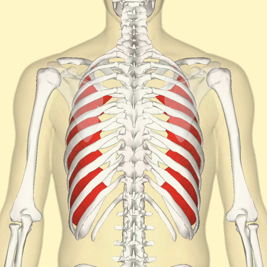
The thoracic cage (rib cage) is the skeletal framework of the thoracic wall, which encloses the thoracic cavity. Abdomen & ribs muscle movements. The rib cage surrounds the lungs and the heart, serving as an important means of bony protection for these vital organs. Rib 2 is thinner and longer than rib 1 and has two articular facets on the head as normal. Various skeletal muscles are attached to the rib cage. Sign up for premium today! The intercostal muscles are the muscles that occupy the 11 intercostal spaces. They are each attached to the ribs. Anterior view of the lungs and ribcage in a transparent female torso stock illustration these pictures of this page are about:human anatomy rib cage muscles. In this lesson i review and critique your assignments on the rib cage. Muscles that move the rib cage attach to the rib cage. Learn about ribs muscle with free interactive flashcards. 1887 human anatomy print of the rib cage and sternum.
On a muscular person when the muscles stretch, we see some of the lower ribs in the front and also in the back. Anterior view of muscle attachments of chest costa. For a gesture drawing, that's good enough. In this lesson i review and critique your assignments on the rib cage. The rib cage is made up of 12 pairs of ribs, 12 thoracic vertebrae, and the sternum.
Intercostal Muscle Strain Physiopedia from www.physio-pedia.com Together these muscles form a column, known as the erector spinae these muscles run up and down over the lower ribs and thorax (the rib cage), and cross to the low. Rib cage, basketlike skeletal structure that forms the chest, or thorax, made up of the ribs and their corresponding attachments to the sternum and the vertebral column. The rib cage is often simplified as an oval shape. The thoracic cage (rib cage) is the skeletal framework of the thoracic wall, which encloses the thoracic cavity. Anatomy of the human body for artists. This is a stereogram, to be viewed in crossview technique. Anterior view of muscle attachments of chest costa. This cage protects vital organs and is essential for creating negative pressure to inflate lungs.
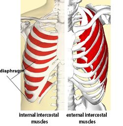
The fibres pass superolaterally to insert into external intercostal muscles internal intercostal muscles.
The rib cage, shaped in a mild cone shape and more flexible than most bone sets, is made up of varying elements such as the thoracic vertebra, 12 equally paired ribs, costal cartilage, and held together anteriorly by the sternum. Another shoulder positioning muscle that can be observed on. This cage protects vital organs and is essential for creating negative pressure to inflate lungs. On a muscular person when the muscles stretch, we see some of the lower ribs in the front and also in the back. Ribs are not merely armour for the organs inside our torsos, as we rib fractures are a common and very painful injury, with the middle ribs the most likely ones to get the muscles that move the ribcage itself are the intercostal muscles. Illustration of thoracic vertebrae showing vertebral body, pedicles, facets, transverse process, rib. During normal breathing, contraction of the major inspiratory muscle, the diaphragm, produces both rib cage expansion and a downward movement of the diaphragm. The ribs joint as follows muscles of the thoracic wall contain those that fill and support the intercostal spaces, those that pass between the sternum and the ribs, and those that cross several. All three groups of muscles support the rib cage. Your rib cage plays an important role in respiration, expanding and contracting as your respiratory muscles, including your diaphragm, work to help you breathe. The rib cage is a primarily protective structure, encircling the heart and lungs. Various skeletal muscles are attached to the rib cage. Structure of a typical rib:
This video includes many structures from thorax and discusses the anatomy of ribs as well as anatomy of rib cage in general rib cage muscles. The intercostal muscles are the muscles that occupy the 11 intercostal spaces.


0 Comments